
Specialty Contact Lenses
Topography Versus Tomography for Irregular Corneas

FSDAVCFEBFEVSDDVFSD
FSDAVCFEBFEVSDDVFSD

FSDAVCFEBFEVSDDVFSD
Why Corneal Imaging Matters
Irregular corneas can make vision blurry, distorted, shadowed, or difficult to correct with standard glasses or soft contact lenses. Conditions such as keratoconus, pellucid marginal degeneration, corneal scarring, or post-surgical changes can alter the shape of the cornea in ways that are not fully explained by a basic prescription.
Corneal imaging helps the doctor understand the structure behind the symptoms. Topography and tomography both provide important information, but they are not exactly the same. Knowing the difference between topography vs tomography irregular corneas can help patients understand why these tests are valuable for diagnosis, monitoring, and specialty contact lens fitting.
Irregular corneas can make vision blurry, distorted, shadowed, or difficult to correct with standard glasses or soft contact lenses. Conditions such as keratoconus, pellucid marginal degeneration, corneal scarring, or post-surgical changes can alter the shape of the cornea in ways that are not fully explained by a basic prescription.
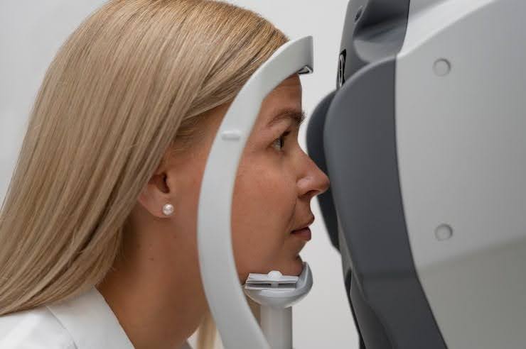
Corneal imaging helps the doctor understand the structure behind the symptoms. Topography and tomography both provide important information, but they are not exactly the same. Knowing the difference between topography vs tomography irregular corneas can help patients understand why these tests are valuable for diagnosis, monitoring, and specialty contact lens fitting.
What Corneal Topography Shows During Exams
A corneal topography scan maps the curvature of the front surface of the cornea. It shows where the cornea is steep, flat, symmetrical, or irregular. This is especially useful when a patient has high astigmatism, distorted vision, keratoconus-like changes, or a history of trouble with regular contact lenses. The map helps the doctor identify patterns that may not be obvious from the prescription alone and provides valuable information about the corneal surface.
For specialty contact lens fitting, topography helps guide the starting lens design. It can reveal whether the cornea has a central cone, inferior steepening, asymmetry, or other irregularities that may affect how a lens should sit. This information is valuable when considering scleral lenses, gas permeable lenses, hybrid lenses, or other custom options to improve comfort and vision.
Topography is also useful for helping patients understand their symptoms. A person may wonder why updated glasses do not provide crisp vision even when the prescription seems correct. Seeing the corneal shape map often makes the explanation easier to understand. The issue may be the irregular surface of the eye rather than simply a prescription problem, allowing the doctor to recommend more appropriate treatment options.
What Corneal Tomography Adds to Diagnosis
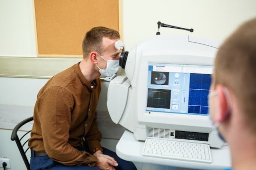
A corneal tomography scan provides a more complete three-dimensional view of the cornea. It evaluates both the front and back surfaces and may also provide information about corneal thickness. This can be especially important when monitoring conditions such as keratoconus or other corneal thinning disorders. The additional data helps doctors evaluate subtle changes that may not be apparent during a routine eye examination.
Tomography can reveal structural changes that may not be fully visible on front-surface topography alone. For patients undergoing irregular cornea testing, this additional information can support diagnosis, progression monitoring, and referral decisions.
It can also help the doctor understand whether a specialty lens fit should be approached with additional caution. Combining tomography with other clinical findings supports more accurate treatment planning, safer specialty contact lens fitting, and better long-term eye health outcomes.
Patients do not need to memorize the technical differences between every scan. The practical point is that topography focuses heavily on the shape of the corneal surface, while tomography adds deeper structural information. Both can help the doctor understand why vision is distorted and what type of lens design may be safest and most effective. Together, these imaging tools provide valuable information for planning specialty contact lens fitting and evaluating complex corneal conditions.
In some cases, one scan may be enough for a particular fitting decision. In more complex cases, both scans may be helpful. A patient with keratoconus progression concerns, unusual corneal shape, prior surgery, or unexplained visual distortion may benefit from multiple forms of imaging. These scans help the doctor avoid guessing and build a lens plan based on better information. The result is a more personalized approach that supports clear vision, improved comfort, and long-term eye health.
How Imaging Improves Lens Fitting
Imaging helps specialty contact lens fitting by showing the shape and structure the lens must work with. A standard lens may not center properly or may fail to correct distortion when the cornea is irregular. Topography and tomography help the doctor choose a better starting point and anticipate fitting challenges before the fitting process begins. These scans provide important details for creating a more customized lens approach.
The scans do not replace trial lens evaluation. The doctor still needs to place lenses on the eye, check comfort, evaluate clearance, assess edge alignment, and confirm vision. However, imaging makes the process more precise because the doctor has a clearer map before beginning the fitting process. This is one reason advanced imaging is valuable during scleral lens fitting Houston patients may undergo, especially when addressing complex corneal conditions.
Why Thickness Information Can Matter
Corneal thickness can be important when the doctor is monitoring conditions that may involve thinning or progression. A corneal tomography scan can provide valuable structural information that helps guide treatment decisions and evaluate the overall health of the cornea.
This does not mean every patient has a serious condition. It simply means the doctor wants to understand the cornea more completely before making recommendations about contact lenses or long-term monitoring. Detailed information supports more informed decisions.
Corneal thickness can be important when the doctor is monitoring conditions that may involve thinning or progression. A corneal tomography scan can provide valuable structural information that helps guide treatment decisions and evaluate the overall health of the cornea.
This does not mean every patient has a serious condition. It simply means the doctor wants to understand the cornea more completely before making recommendations about contact lenses or long-term monitoring. Detailed information supports more informed decisions.
For specialty lens patients, thickness and shape data may help guide follow-up timing, lens expectations, and whether additional medical evaluation is appropriate. More complete information often leads to safer and more customized care. These measurements help create a personalized approach for better vision outcomes and long-term eye health.
Why Better Imaging Leads to Better Decisions
Topography and tomography are especially useful because irregular corneas can behave differently from one patient to another. Two people may have similar glasses prescriptions but very different corneal shapes. One patient may do well with a relatively simple lens design, while another may require irregular cornea contact lenses, a scleral lens, or multiple fitting adjustments. Imaging helps explain those differences and allows the doctor to understand the unique characteristics of each cornea before beginning the fitting process at Kleinwood Vision.
For patients, these scans can also reduce frustration. Many people with irregular corneas have spent years being told they have astigmatism, dry eye, or a difficult prescription. When imaging reveals the true shape issue, the path forward becomes clearer. The doctor can explain whether the goal is sharper vision, improved comfort, safer lens clearance, or monitoring for future changes that may affect eye health.
The most effective specialty lens fit usually comes from combining imaging with real-world testing. Topography and tomography provide the map, but the lens still has to prove itself on the eye. Patient feedback about comfort, fogging, vision stability, redness, dryness, and wear time remains essential throughout the specialty contact lens fitting process.
Together, imaging and specialty contact lens fitting help create a more customized and safer lens plan. By using detailed corneal information along with trial lens evaluation, doctors can make better decisions about lens design, adjustments, and long-term care. This approach supports clearer vision, better comfort, and successful specialty lens wear for patients with complex corneal needs while improving confidence in the overall treatment process.
Questions to Ask About Your Scans
Ask what your corneal topography scan shows about the shape of your cornea. Understanding whether irregular curvature is affecting your vision can make it easier to understand why certain lens options are being recommended. This information helps explain how corneal shape influences specialty contact lens fitting and supports better treatment decisions for clearer vision during comprehensive eye exams.
Ask whether a corneal tomography scan is needed to evaluate corneal thickness or deeper structural changes. This can be especially important if keratoconus, pellucid marginal degeneration, or progression concerns are part of the discussion. Detailed imaging provides important information about corneal health and helps guide appropriate care.
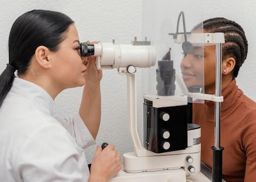
Ask what your corneal topography scan shows about the shape of your cornea. Understanding whether irregular curvature is affecting your vision can make it easier to understand why certain lens options are being recommended. This information helps explain how corneal shape influences specialty contact lens fitting and supports better treatment decisions for clearer vision during comprehensive eye exams.
Ask whether a corneal tomography scan is needed to evaluate corneal thickness or deeper structural changes. This can be especially important if keratoconus, pellucid marginal degeneration, or progression concerns are part of the discussion. Detailed imaging provides important information about corneal health and helps guide appropriate care.
Finally, ask how the imaging results will guide your lens fit. The answer should connect the scans to lens design, expected comfort, vision goals, and future follow-up visits. Understanding this process helps patients make confident decisions about customized eye care.
Use Better Imaging for Better Fitting
If you have an irregular cornea, distorted vision, keratoconus, pellucid marginal degeneration, or repeated contact lens fitting problems, corneal imaging can help explain what is happening. Bring previous records if available, especially older scans, prescriptions, or lens information, so the doctor can compare changes over time and make more informed decisions. This information supports accurate evaluation and helps create a personalized care plan.
Kleinwood Vision uses advanced imaging and comprehensive eye exams to evaluate irregular corneas and support customized lens care. Whether you need specialty contact lens fitting, advanced irregular cornea testing, or guidance through the scleral lens fitting Houston process, our team can help. Contact us to learn how detailed imaging and personalized lens solutions may improve your vision, comfort, and long-term eye health. These services are designed to support clearer vision and better outcomes.
Contact Info
Hours of Operation
Mon - Fri | 9:00 AM - 5:00 PM
Sat - Sun | Closed
Holiday Hours: We are closed for the following holidays: New Years Day, Memorial Day, Independence Day, Labor Day, Thanksgiving Day, Christmas Day
The information provided on this website is for general informational purposes only and does not constitute professional medical advice, diagnosis, or treatment. Always seek the guidance of a licensed eye care professional or qualified health provider with any questions you may have regarding a medical condition or vision concern. Results from eye care services may vary by individual.
© 2026 Kleinwood Vision. All rights Reserved.