
Retinal Care
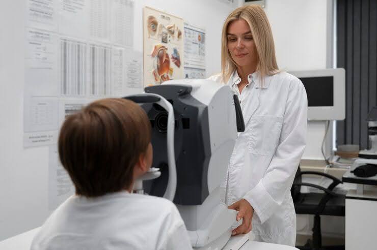
Retinal Imaging Steps for High Myopia

FSDAVCFEBFEVSDDVFSD
FSDAVCFEBFEVSDDVFSD

FSDAVCFEBFEVSDDVFSD
Why High Myopia Needs Watchful Care
An elongated eye in high myopia stretches delicate retinal tissues, increasing high myopia retinal risks such as tears, detachments, and the need for myopic maculopathy screening. Since early retinal changes can occur without symptoms, scheduled retinal imaging works like a preventive smoke alarm—alerting doctors to issues before vision is affected.
Families should plan a clear prevention pathway that includes baseline wide-field imaging, regular follow-up reviews, and quick access to care if alarming symptoms appear. The goal is straightforward: detect small changes early and take action to protect long-term vision in high myopia, reducing the risk of serious retinal complications.
Imaging Tools That See More
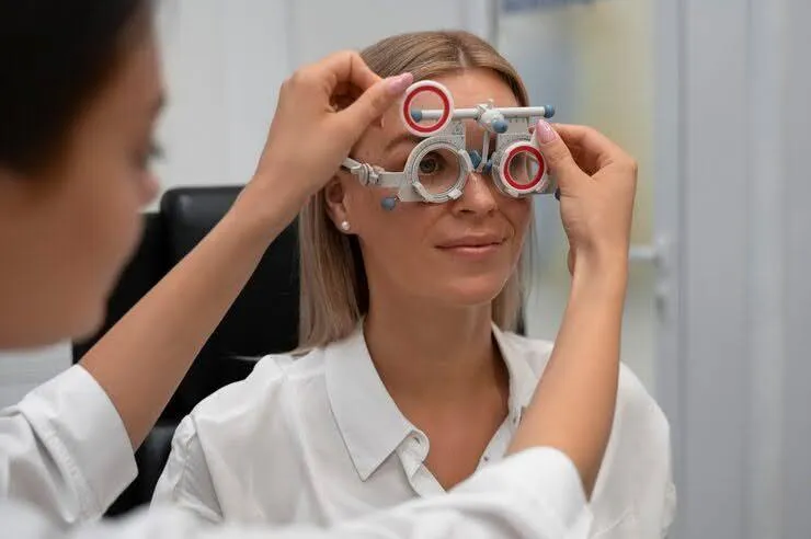
Wide-field retinal imaging captures the far-peripheral retina—where many retinal tear detection events begin—going beyond what standard views can reveal. This technology allows clinicians to spot early warning signs that might otherwise go unnoticed. An OCT scan eyes layer by layer, detecting subtle macular changes that are invisible in surface exams. Meanwhile, a dilated eye exam enables direct inspection of every retinal quadrant in real time. These tools work together: wide-field provides a broad map, OCT offers detailed depth, and dilation allows hands-on examination.
Clinics archive these imaging results to create a visual history for each patient. Comparing images side by side over time helps identify meaningful changes from your personal baseline—not just one-off findings. This historical view guides decisions about whether intervention or referral is necessary, supporting safer and more personalized high myopia retinal risk management strategies.
Clinicians focus on trends like new lesions, expansion, or retinal thinning rather than reacting to isolated abnormalities. This layered, comprehensive approach builds confidence in clinical decisions, ensures more stable monitoring over time, and may help avoid unnecessary treatments. For patients with high myopia, it offers a more precise, thoughtful way to protect long-term retinal health.
Understanding Your Report: What to Know

Your report may mention findings like lattice degeneration, early posterior vitreous detachment, or posterior staphyloma. Each of these will be labeled as “monitor,” “treat,” or “urgent,” helping guide your next steps. Ask your Houston optometrist to show last year’s image side-by-side with today’s—seeing visual trends over time makes potential risks and progression in high myopia much easier to understand and track accurately.
You should leave the visit with clear, plain-language instructions outlining your next steps.
This includes your recommended return interval, any activity precautions based on your findings, and which symptoms should be considered red flags requiring same-day attention. Knowing exactly what to watch for between visits helps protect your vision and supports safe, long-term management. When patients understand their reports and plans, it leads to better outcomes in managing high myopia and related retinal health concerns.
Always memorize red-flag symptoms: sudden new floaters, brief flashes of light, a curtain-like shadow across your vision, or sudden loss of peripheral vision. These signs may indicate serious retinal issues, especially in individuals with high myopia. If any of these symptoms occur, call the clinic the same day—earlier is always safer. Prompt reporting allows your eye care provider at Kleinwood Vision to evaluate and address potential problems before they progress, helping to protect your long-term vision.
Keep your archived images accessible through your patient portal or store them securely in your personal records. These scans serve as a valuable baseline for future comparisons. If you ever relocate or require urgent eye care while traveling, having access to your prior retinal scans can significantly speed up care and avoid unnecessary or redundant imaging. Being prepared ensures continuity of care and allows any provider to make informed decisions quickly, especially when managing high myopia retinal risks.
Cadence, Lifestyle, and Prevention Strategies Guide
How often you receive retinal imaging depends on your individual risk level and past findings. Eyes with higher-risk features—such as lattice degeneration or posterior staphyloma—are imaged more frequently, while stable cases may extend to yearly intervals. Alongside imaging, support eye health with UV-blocking eyewear, safe sports practices to prevent trauma, and strong pediatric myopia control strategies aimed at slowing axial growth.
Consistency outperforms extremes when managing high myopia. It’s not about doing everything all at once, but about maintaining steady, protective habits over time. Combining routine imaging with daily lifestyle choices builds a strong defense against progression. Scheduled reviews help detect early changes, while consistent care—at home and in clinic—protects long-term vision. Prevention isn’t one action; it’s a pattern of care that supports healthy eyes through each stage of high myopia management.
Visit Prep and Record-Keeping Checklist Guide
For your visit, bring your current glasses and a detailed timeline of any symptoms such as floaters, flashes, or visual distortions. This information helps your eye care provider assess changes more accurately and detect potential high myopia complications early.
Ask ahead if a dilated eye exam is planned, as your vision may be blurry afterward. If so, arrange safe transportation to and from the clinic. Being prepared ensures you get the full benefit of the visit without added stress.
Request copies of your retinal images or ask about patient portal access. Keeping your own record supports second opinions, urgent referrals, and ongoing care—especially if you relocate or need care while traveling. Organized records help streamline communication and improve long-term high myopia management.
Request copies of your retinal images or ask about patient portal access. Keeping your own record supports second opinions, urgent referrals, and ongoing care—especially if you relocate or need care while traveling. Organized records help streamline communication and improve long-term high myopia management.
Escalation and Referral Pathways for Care
If imaging or an exam suggests a retinal tear, an urgent referral to a retinal specialist is essential. Retinal tears can lead to retinal detachment if left untreated, which can cause permanent vision loss. Treatment options depend on the tear’s size and location and may include laser barricade therapy, which seals the tear, or vitrectomy surgery, which removes vitreous traction to prevent further damage. Early intervention is critical to protect your vision and prevent serious complications.
For macular changes or early myopic degeneration, Optical Coherence Tomography (OCT) is used to monitor the retina’s layers for fluid accumulation, vitreous traction, or thinning. The management approach varies based on severity; some cases require close monitoring with regular imaging, while others might need intravitreal therapy, where medication is injected into the eye to control disease progression. Timely detection through OCT enables tailored treatment to preserve central vision.
Your Houston optometrist typically coordinates referrals and follow-up care with retinal specialists. After the specialist stabilizes the eye, your optometrist resumes long-term surveillance to monitor ongoing retinal health. They will document your next recommended return interval and review red-flag symptoms to watch for between visits, such as new floaters, flashes, or vision changes.
Clear escalation and referral pathways ease patient and family anxiety and save time when vision matters most. Knowing when to seek urgent care and having coordinated support helps ensure safe, effective management of retinal risks in high myopia, safeguarding your vision for the long term.
Patient FAQs About Retinal Imaging Explained
Do wide-field photos replace dilation? Not always—they serve different but complementary roles. Wide-field photos provide a quick overview of the retina, while dilation allows a more detailed exam of eye health. Both are important in thorough eye care. Does OCT hurt? No, OCT is a quick, non-contact scan of your retina that is painless and safe.
Is imaging covered by insurance? Sometimes—medical indications typically qualify, but coverage varies by plan. It’s best to check your insurance policy ahead of time.

Do wide-field photos replace dilation? Not always—they serve different but complementary roles. Wide-field photos provide a quick overview of the retina, while dilation allows a more detailed exam of eye health. Both are important in thorough eye care. Does OCT hurt? No, OCT is a quick, non-contact scan of your retina that is painless and safe.
Is imaging covered by insurance? Sometimes—medical indications typically qualify, but coverage varies by plan. It’s best to check your insurance policy ahead of time. How fast are results? Many imaging results are available immediately, allowing your doctor to discuss findings during your visit. Complex or subtle issues may require specialist review, which can take longer.
How fast are results? Many imaging results are available immediately, allowing your doctor to discuss findings during your visit. Complex or subtle issues may require specialist review, which can take longer.
Is pregnancy a concern? Your doctor will adjust dilation timing and comfort as needed during pregnancy to ensure safety. Always inform your provider if you are pregnant or suspect pregnancy to receive appropriate care.
Is pregnancy a concern? Your doctor will adjust dilation timing and comfort as needed during pregnancy to ensure safety. Always inform your provider if you are pregnant or suspect pregnancy to receive appropriate care.
Your Protection Plan at Home
Monitor for any new floaters, flashes, or curtain-like shadows in your vision, and if you notice any, call the clinic the same day. These symptoms are important red flags that require prompt evaluation to prevent serious retinal complications. Protect your eyes by wearing UV-blocking sunglasses when outdoors, keeping your prescriptions updated, and following your myopia-management plan consistently. These steps help slow axial growth and reduce high myopia retinal risks. If you have concerns between visits, contact us for guidance.
At the end of each visit, make sure to schedule your next imaging appointment. Regular, steady surveillance combined with healthy habits offers the best protection against progression. Consistent monitoring through scheduled imaging allows early detection of changes, enabling timely intervention. This proactive approach helps preserve your long-term retinal health and vision, ensuring that you stay ahead of any complications related to high myopia.
Contact Info
Hours of Operation
Mon - Fri | 9:00 AM - 5:00 PM
Sat - Sun | Closed
Holiday Hours: We are closed for the following holidays: New Years Day, Memorial Day, Independence Day, Labor Day, Thanksgiving Day, Christmas Day
The information provided on this website is for general informational purposes only and does not constitute professional medical advice, diagnosis, or treatment. Always seek the guidance of a licensed eye care professional or qualified health provider with any questions you may have regarding a medical condition or vision concern. Results from eye care services may vary by individual.
© 2026 Kleinwood Vision. All rights Reserved.