
Comprehensive Eye Exams
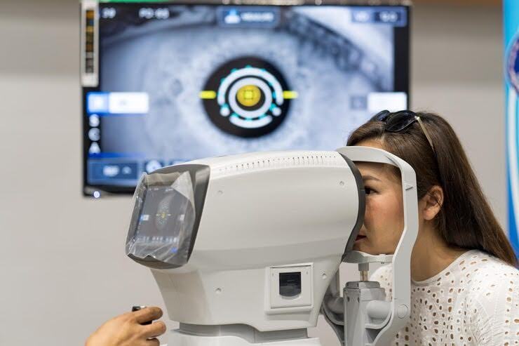
Optomap Retinal Imaging Explained

FSDAVCFEBFEVSDDVFSD
FSDAVCFEBFEVSDDVFSD

FSDAVCFEBFEVSDDVFSD
What Wide-Field Retinal Imaging Does
Optomap retinal imaging creates a wide-field view of the retina, the light-sensitive tissue lining the back of the eye. This helps your eye doctor document and review areas that are difficult to appreciate fully with basic front-of-eye testing alone. The image becomes part of your record, which can be useful for comparing changes over time and explaining findings more clearly during the exam.
For many patients, this makes the eye health discussion easier to understand. Instead of hearing only a verbal description, you can see an image of your retina and talk through why certain areas matter. Wide-field imaging can support screening for retinal changes, help monitor known findings, and add another layer of documentation during a comprehensive eye exam in Houston.
What Conditions It Can Help Reveal
Retinal imaging can help identify or document a variety of findings, including retinal holes, tears, areas of degeneration, pigment changes, diabetic eye changes, hypertensive changes, nevi, and other abnormalities that deserve monitoring or referral. It is also useful for establishing a baseline in patients who are at higher risk for retinal disease or who have findings that should be followed from year to year.
This wide-field view is especially helpful because the retina extends far beyond the small area patients think of as the “back of the eye.” Peripheral findings can matter even when central vision feels normal. Imaging may reveal changes before a patient notices symptoms, which is one reason comprehensive eye health screening remains important even for people who believe they “see fine.”
At the same time, retinal imaging should be understood as a powerful tool, not a simplistic replacement for clinical judgment. Some findings need closer direct examination, correlation with symptoms, or additional testing to understand their significance. The image helps guide care and improve documentation, but your optometrist still decides whether the retina has been assessed completely enough or whether dilation is needed to answer the clinical question safely.
Who Benefits Most from Optomap Imaging
Many patients can benefit from retinal imaging, but it is especially valuable for those with diabetes, high myopia, a family history of retinal disease, past retinal findings, flashes or floaters history, or any condition that increases concern about retinal health. It can also be helpful for children and adults who want a more visual explanation of their eye health or who appreciate having a documented baseline for future comparison.
Patients wearing strong prescriptions often benefit because retinal risk can be higher in more myopic eyes, making careful peripheral evaluation more important.
Imaging is also useful when the doctor wants to show a patient why a finding should be monitored rather than leaving the conversation abstract. Seeing the retina can improve understanding, follow-through, and confidence in the care plan.
One of the strengths of Optomap imaging is that it adds documentation you can return to later. If a small retinal spot, lattice degeneration area, or pigment change is being watched over time, having prior images can help your doctor compare whether it looks stable or whether it appears to be changing. That kind of side-by-side review is useful for long-term monitoring and can make follow-up recommendations feel more concrete. Patients also tend to remember the conversation better when they can see what the doctor is talking about. Imaging does not just help the clinician; it often improves patient understanding in a practical way that makes yearly care feel more meaningful.
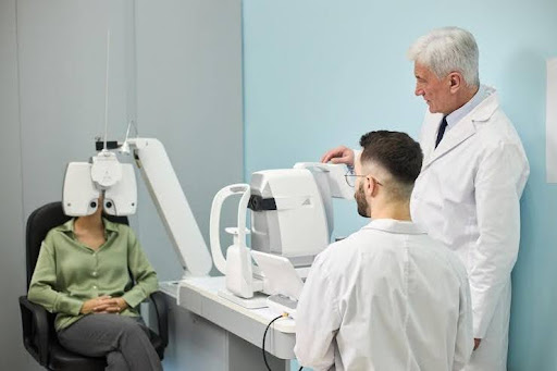
Why Dilation Can Still Be Necessary
Even with excellent retinal imaging, dilation still matters in some situations. If you have new flashes, floaters, visual field changes, suspicious retinal findings, media opacity, or symptoms that raise concern for a peripheral retinal problem, your doctor may still recommend dilation to examine the retina more completely and directly. Imaging is valuable, but some clinical situations require the broadest possible view and the flexibility of a full dilated assessment.
This is why retinal imaging should not be framed as an all-or-nothing substitute for dilation. In many routine cases, imaging adds substantial value and may reduce uncertainty about what the retina looks like at that visit. But if the doctor needs a more thorough view or wants to evaluate a finding more carefully, dilation remains an important part of comprehensive care. The right approach depends on the patient’s history, symptoms, and exam findings that day.
Why Seeing the Image Helps Patients
Patients often understand their eye health better when they can actually see a retinal image instead of hearing a technical description. The conversation becomes more concrete and easier to remember, helping patients feel more involved in their own care and decisions.
This is especially useful when the doctor is monitoring something over time. It helps patients understand why a recheck matters and why “everything looks stable” can be meaningful even if they do not feel anything different or notice obvious changes in vision.

A visual record also supports better questions. Once patients can see the image, they often become more engaged in the exam and more confident about the next step in care, leading to clearer communication and improved understanding.
Imaging and Dilation Work Together
Optomap retinal imaging is most useful when patients understand both its strengths and its limits. It provides your eye doctor a broad documented view of retinal health, helps monitor changes over time, and can reveal important findings even in people without obvious symptoms. It also improves communication because patients can see what is being discussed rather than trying to imagine it. For people with higher retinal risk or those who value detailed baseline documentation, that can make comprehensive eye care feel much more informative and precise.
At the same time, good care depends on choosing the right tools for the specific situation. Imaging is not a shortcut around thoughtful examination, and dilation is not outdated just because imaging exists. Both approaches remain important in evaluating retinal health thoroughly and safely during a complete eye exam.
They serve related but different purposes. In many visits, wide-field imaging like Optomap retinal imaging adds excellent information by capturing a large view of the retina quickly and comfortably. In other situations, dilation remains necessary to allow a closer and more detailed inspection of subtle retinal structures that imaging may not fully show. Each method contributes unique clinical value depending on the patient’s needs and risk factors.
The goal is not to force one method to replace the other. It is to use each in the way that best protects retinal health and helps patients understand what their exam actually showed. When combined appropriately, these tools provide a more complete and reliable picture, supporting early detection, better monitoring, and more informed decisions for long-term eye health and visual protection.
Questions to Ask During the Exam
Ask what the imaging shows today and whether there is anything your doctor wants to monitor over time. That turns the image into a useful part of the conversation instead of just another test and helps you understand how it relates to your current eye health and future care plan.
It is also helpful to ask whether your personal history makes wide-field retinal imaging especially valuable. Patients with myopia, diabetes, or prior retinal findings may benefit from understanding why imaging is being emphasized in their case and how it supports long-term monitoring and early detection of changes.
Finally, ask whether dilation is still recommended and why. Knowing why your doctor wants more direct examination makes the decision feel more purposeful and easier to accept, while also helping you understand how dilation and imaging work together to give a more complete and accurate view of retinal health.
Your Next Steps Before Your Visit
Before your eye exam, think about whether you have had flashes, floaters, retinal concerns, diabetes, high prescriptions, or previous advice to monitor a retinal finding. Bring your current glasses and be ready to mention any family history of retinal disease if you know it. These details help your eye doctor decide how important imaging and dilation may be during your visit and ensure a more complete evaluation of your retinal health.
If you want a clearer view of your retinal health, Kleinwood Vision can explain how Optomap retinal imaging fits into a comprehensive eye exam and whether dilation still makes sense based on your symptoms and risk factors. Understanding both tools helps you see how they complement each other, supporting better decisions about your eye health screening, early detection, and long-term care planning for more confident and informed vision care. Contact us today to schedule your appointment.
Contact Info
Hours of Operation
Mon - Fri | 9:00 AM - 5:00 PM
Sat - Sun | Closed
Holiday Hours: We are closed for the following holidays: New Years Day, Memorial Day, Independence Day, Labor Day, Thanksgiving Day, Christmas Day
The information provided on this website is for general informational purposes only and does not constitute professional medical advice, diagnosis, or treatment. Always seek the guidance of a licensed eye care professional or qualified health provider with any questions you may have regarding a medical condition or vision concern. Results from eye care services may vary by individual.
© 2026 Kleinwood Vision. All rights Reserved.