
Eye Exams
Understanding Modern Eye Imaging Tests

FSDAVCFEBFEVSDDVFSD

FSDAVCFEBFEVSDDVFSD

FSDAVCFEBFEVSDDVFSD
Why Eye Imaging Is Part of Modern Exams
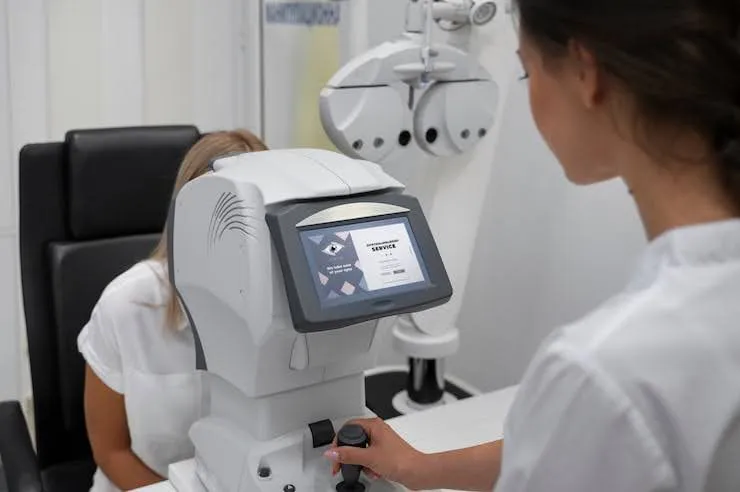
Modern eye care relies on advanced imaging to evaluate structures that cannot be fully assessed with vision testing alone. During a comprehensive eye exam Houston patients receive, imaging provides detailed views of the retina and optic nerve, creating baseline records for future comparison. These images help detect subtle structural changes that may not cause symptoms right away, improving diagnostic accuracy and supporting long-term monitoring.
Many eye diseases—including glaucoma, macular degeneration, and diabetic retinopathy—develop silently. Imaging allows earlier identification, often before vision changes occur. When patients understand how scans support prevention, they feel more confident in their care. At Kleinwood Vision, imaging supports lifelong visual health.
OCT vs Retinal Photos vs Wide-Field Scans
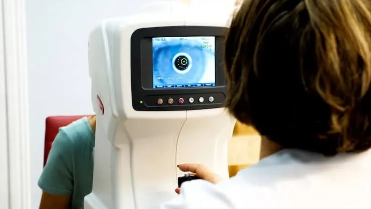
When patients want the OCT eye test explained, it helps to think of it as a cross-sectional scan of the retina. Optical Coherence Tomography (OCT) measures and maps the layers of retinal tissue and the optic nerve with microscopic precision. It detects swelling, thinning, or early structural damage that may not appear in photographs, making it essential for eye imaging for glaucoma and macular evaluation. Because OCT captures detailed structural data, it allows doctors to monitor subtle progression over time and adjust treatment plans with greater confidence and accuracy.
A retinal photography exam captures high-resolution surface images of the retina. These photos document blood vessels, the optic nerve head, and pigment patterns. They are useful for tracking diabetic changes, vascular abnormalities, and establishing a clear visual baseline for future comparisons during routine exams and ongoing disease management.
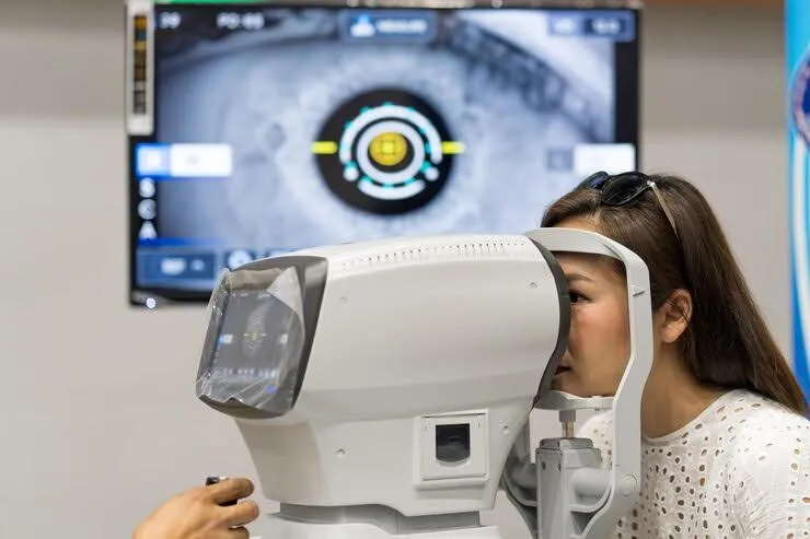
Wide-field retinal imaging expands the visible area to include the peripheral retina. This broader view helps detect tears, holes, lattice degeneration, and peripheral diabetic changes that standard images might miss. Because each test answers different clinical questions, doctors often combine multiple imaging methods for a more complete and proactive assessment.
What Patients Experience During Each Test
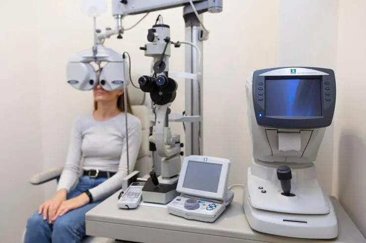
Most imaging tests are quick, non-contact, and painless. Patients simply look at a fixation target while the device captures images in seconds. OCT may require several scans per eye, making it important to keep your eyes steady throughout the process to ensure the most precise results. Retinal photography uses a brief, bright flash, while wide-field retinal imaging can feel slightly more intense due to its broader illumination, which captures both central and peripheral areas of the retina.
Some imaging can be completed without dilation, though dilation may still be recommended depending on pupil size and clinical findings.
Knowing what to expect reduces anxiety and improves patient cooperation during the exam. Staying relaxed and following instructions carefully helps produce clearer images and more accurate results, supporting thorough evaluation, earlier detection of eye conditions, and better-informed decisions about your long-term eye health during your visit.
Imaging provides the most value when results are compared over time. Baseline scans allow doctors to detect small but meaningful changes, such as early optic nerve thinning or subtle macular fluid. Identifying these changes early can guide treatment decisions before noticeable vision loss occurs, supporting proactive management of conditions like glaucoma, macular degeneration, or diabetic retinopathy. Regular follow-up imaging ensures that any progression is tracked accurately, giving patients and doctors the information needed to make timely adjustments to treatment plans and maintain long-term eye health.
Clear imaging depends on steady fixation and healthy tear film quality. Blinking between scans and following instructions carefully helps prevent blur and ensures sharp, reliable images. If images are unclear due to dry eye, small pupils, or other factors, repeat captures may be necessary. Consistent, high-quality imaging reduces the risk of missing early disease signs and strengthens long-term monitoring accuracy, allowing for earlier detection and more precise evaluation of changes in retinal and optic nerve structure.
When Each Imaging Type Is Most Useful
OCT is commonly used for monitoring glaucoma, evaluating macular degeneration, and assessing diabetic macular edema. Because it measures retinal thickness with microscopic precision, OCT plays a key role in eye imaging for glaucoma and tracking disease progression before vision is noticeably affected. Regular OCT scans allow doctors to detect subtle changes early, guiding treatment decisions and helping preserve long-term visual health.
A retinal photography exam documents baseline retinal appearance and monitors blood vessel or pigment changes over time. Wide-field retinal imaging is especially valuable for patients with high myopia, new flashes or floaters, or those undergoing a comprehensive diabetic eye exam that includes peripheral screening. Your doctor selects the most appropriate imaging method based on your symptoms, risk profile, and exam findings, ensuring a personalized approach to eye care and accurate monitoring for any potential changes in retinal or optic nerve structure.
Imaging Limits and Why Exams Still Matter
Imaging does not replace a full eye examination. Certain conditions require dilation, visual field testing, or direct clinical evaluation to confirm findings and ensure a comprehensive assessment. Relying solely on images may miss important details that only a complete exam can detect.
Images may also be affected by cataracts, dry eye, or difficulty maintaining fixation. Blurry scans sometimes need to be repeated to ensure accuracy and reliability. Clear, high-quality imaging is essential for meaningful results and effective monitoring over time.
Patients should view imaging as one part of a complete diagnostic picture. When multiple tests are recommended, it is because each contributes unique information that strengthens overall decision-making. Using imaging alongside clinical evaluation helps protect long-term eye health and provides the most accurate assessment of retinal and optic nerve conditions.
How Imaging Protects Long-Term Vision
One of the greatest advantages of modern imaging is early detection. OCT can reveal structural optic nerve changes associated with glaucoma before noticeable vision decline occurs. When the OCT eye test explained includes its ability to measure microscopic tissue changes, patients often feel reassured knowing potential problems can be identified early. Detecting these subtle changes allows doctors to monitor progression carefully and take timely action, supporting proactive eye care and reducing the risk of vision loss.
For patients managing diabetes, diabetic eye exam imaging plays a critical role in spotting microvascular changes long before symptoms develop. A retinal photography exam documents visible vessel changes, while wide-field retinal imaging captures peripheral areas where damage may first appear. Early identification supports prompt referral or treatment, helping prevent advanced complications and preserving long-term retinal health.
Macular conditions also benefit from detailed imaging. OCT detects fluid buildup, subtle swelling, or disruptions within retinal layers that guide treatment decisions. Even when results appear normal, baseline scans provide valuable comparison data for the future. This allows doctors to track changes over time, ensuring that subtle developments are addressed before they affect vision.
Patients who include imaging as part of their regular comprehensive eye exam Houston visits are more likely to catch issues early. Ongoing monitoring at Kleinwood Vision supports precise tracking, earlier intervention, and greater confidence in long-term visual stability. By combining OCT, retinal photography, and wide-field imaging, patients receive a complete evaluation that protects both retinal and optic nerve health for years to come.
Common Imaging Questions Patients Ask
Many patients ask whether imaging replaces dilation. While advanced scans can sometimes reduce the need for dilation, it may still be necessary for a complete evaluation of the retina. Imaging provides valuable information, but a full dilated exam ensures that subtle or peripheral issues are not missed.
Others wonder whether flashes always indicate a retinal tear. Although wide-field retinal imaging can help detect peripheral problems, only a comprehensive examination confirms the cause. Similarly, some people ask if OCT is like an MRI. OCT uses light waves—not radiation or magnets—to measure retinal layers and cannot directly diagnose brain conditions, making it very different from an MRI.
Questions about frequency are also common. Imaging schedules depend on individual risk factors such as diabetes, high myopia, or family history of glaucoma. If you are unsure which scans are appropriate for you, Contact us for personalized guidance.
Personalized Plan for Your Retinal Imaging
Ask your eye doctor which imaging tests you need and why. Request baseline copies when available and follow recommendations for dilation or repeat scans. Tracking changes over time is one of the most effective ways to protect your vision, as it allows your eye care team to identify subtle shifts before they become serious problems. Consistent monitoring helps detect early signs of conditions like glaucoma, diabetic retinopathy, or macular degeneration, ensuring timely intervention and better long-term outcomes for your eyes.
If you experience new flashes, floaters, or sudden blurred vision, Contact us promptly—imaging may become urgent rather than routine. Combining advanced diagnostic scans with regular exams at Kleinwood Vision provides one of the strongest strategies for safeguarding lifelong eye health. Proactive imaging and routine exams work together to preserve vision and maintain overall eye wellness.
Contact Info
Hours of Operation
Mon - Fri | 9:00 AM - 5:00 PM
Sat - Sun | Closed
Holiday Hours: We are closed for the following holidays: New Years Day, Memorial Day, Independence Day, Labor Day, Thanksgiving Day, Christmas Day
The information provided on this website is for general informational purposes only and does not constitute professional medical advice, diagnosis, or treatment. Always seek the guidance of a licensed eye care professional or qualified health provider with any questions you may have regarding a medical condition or vision concern. Results from eye care services may vary by individual.
© 2026 Kleinwood Vision. All rights Reserved.